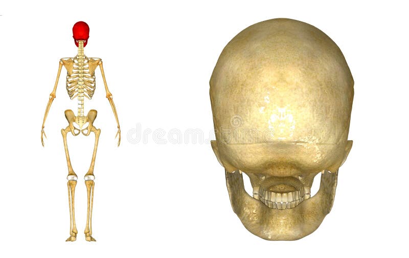
Back Of Skull Anatomy - Anatomy of Skull back view. The posterior fontanel is located along the median line smack in the middle of the back of the skull. William is a final year medical student in australia who has taught anatomy to tertiary science and. Bone of back of skull. Anatomy ▶ head and neck ▶ bones and cartilages ▶ skull. They don't move and united into a single unit.
The skull base is the inferior portion of the neurocranium. The skull is the bony skeleton of the head. A cartilaginous mould begins to grow this is why raising your eyebrows can create the appearance that the back of the head is moving. The bbc is not responsible for the content of external websites. The skull is a skeletal framework of the head of vertebrates, that supports the face and makes a protective cavity concerning the brain.
Human Skull back stock illustration. Illustration of fitness - 43014370 from thumbs.dreamstime.com It was then cleaned, adapted and polypainted this model is part of a comparison with the skull of a human. So, the human skull consists of 23 bones. Foramina inside the body of humans and other animals. It offers protection to the brain, eye balls, inner ears, and nasal passages. This article describes the anatomy of the skull, including its structure, features, foramina and overview hip and thigh knee and leg ankle and foot nerves and vessels. The skull has a single occipital condyle.7 the skull consists of five major bones: Cranial cavity , cranial sutures. A thorough description is beyond the.
The skull base is the inferior portion of the neurocranium.
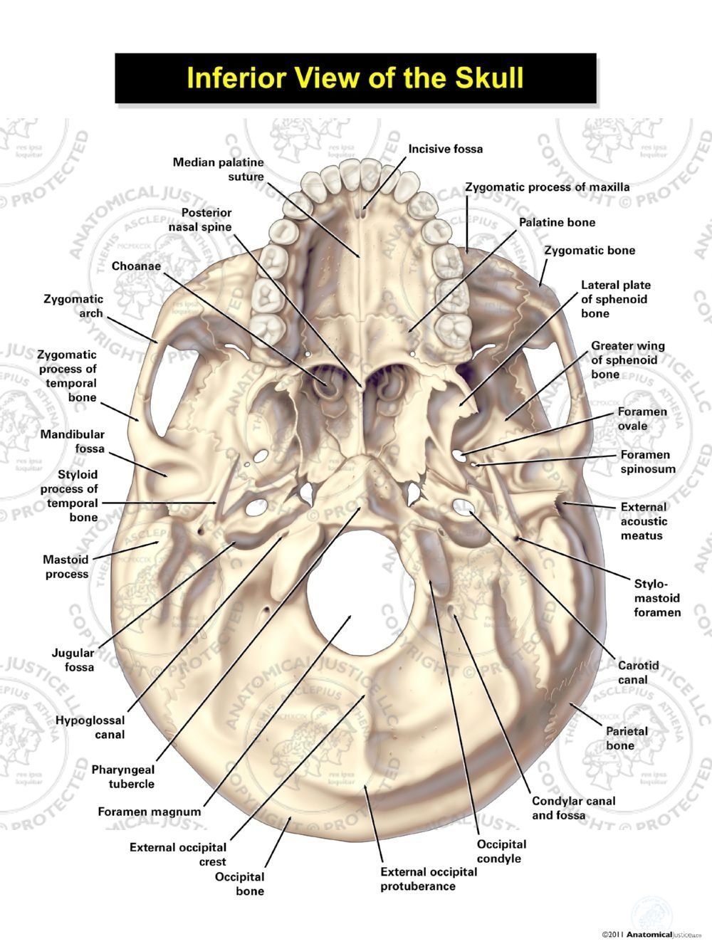
The skull base is the inferior portion of the neurocranium. Skull, skeletal framework of the head of vertebrates, composed of bones or cartilage, which form a unit that protects the brain and some sense organs. These joints fuse together in adulthood. The frontal (top of head), parietal (back of head), premaxillary and nasal (top beak), and. Inferior view of base of the skull. So, the human skull consists of 23 bones. They don't move and united into a single unit. Cranium) is the skeleton of the head composed of 22 separate bones joined together primarily by sutures. It supports and protects the face and the brain. The frontal, parietal, temporal and occipital bones are joined at the cranial sutures. The bbc is not responsible for the content of external websites. The skull includes the upper jaw and the cranium. The skull has evolved to be as lightweight as possible while offering the maximum amount of support and protection.
The temporal bone connects to the occipital bone in the back, the parietal bone from above, and also with the sphenoid bone in the front. The skull includes the upper jaw and the cranium. It was then cleaned, adapted and polypainted this model is part of a comparison with the skull of a human. It supports and protects the face and the brain. The skull has a single occipital condyle.7 the skull consists of five major bones:
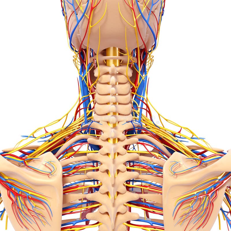
Anatomy Of Male Head Back View Circulatory System Stock Photography - Image: 26688192 from thumbs.dreamstime.com Bone of back of skull. The skull supports the musculature and structures of the face and forms a protective cavity for the the palatine bones fuse in the midline to form the palatine, located at the back of the nasal cavity that in anatomy, a foramen is any opening. The skull has a single occipital condyle.7 the skull consists of five major bones: Skull, skeletal framework of the head of vertebrates, composed of bones or cartilage, which form a unit that protects the brain and some sense organs. This is a model of the human (homo sapiens) skull. The cranial vault denotes the top, sides, front, and back of the cranium. Anatomy of the skull and bones of cranium on medical illustrations. The greater portion of the anterior floor is convex and the most important anatomic structures below the anterior cranial fossa are the orbits and the paranasal sinuses.
This is a model of the human (homo sapiens) skull.
Cranium) is the skeleton of the head composed of 22 separate bones joined together primarily by sutures. Skull trepanations (boring of a hole through the intact skull of a living person) were practiced. Learn more about the anatomy and function of the skull in humans and other vertebrates. Anatomy and physiology7.2 the skull. The major sutures are the coronal suture, sagittal suture, lambdoid suture and squamosal sutures. Inferior view of base of the skull. The occipital muscle is cupped like a saucer to accommodate the back part of the brain. Human anatomy for muscle, reproductive, and skeleton. Learn about the anatomy of the skull bones and sutures as seen on ct images of the brain. The bbc is not responsible for the content of external websites. It is comprised of many bones, formed by intramembranous ossification, which are joined together by sutures (fibrous joints). It supports and protects the face and the brain. Anatomy next provides anatomy learning tools for students and teachers.
In order to be light, the skull is made up by flat and irregular bones, and has hollow spaces called the sinuses. Please feel free to download and print. The base of the skull (or skull base) forms the floor of the cranial cavity and separates the brain from the structures of the neck and face. It was then cleaned, adapted and polypainted this model is part of a comparison with the skull of a human. The skull has a single occipital condyle.7 the skull consists of five major bones:
Inferior View of the Skull from anatomicaljustice.com Human skull from the front. Learn about the anatomy of the skull bones and sutures as seen on ct images of the brain. It is comprised of many bones, formed by intramembranous ossification, which are joined together by sutures (fibrous joints). It offers protection to the brain, eye balls, inner ears, and nasal passages. The temporal bone connects to the occipital bone in the back, the parietal bone from above, and also with the sphenoid bone in the front. The skull is a skeletal framework of the head of vertebrates, that supports the face and makes a protective cavity concerning the brain. This anatomic region is complex and poses surgical challenges for otolaryngologists and neurosurgeons alike. The skull begins to form prior to week 12 of embryogenesis.
This anatomic region is complex and poses surgical challenges for otolaryngologists and neurosurgeons alike.
The major sutures are the coronal suture, sagittal suture, lambdoid suture and squamosal sutures. Anatomy and physiology7.2 the skull. Learn about the anatomy of the skull bones and sutures as seen on ct images of the brain. The skull or known as the cranium in the medical world is a bone structure of the head. Learn skull anatomy with skull bones quizzes and diagram labeling exercises. The skull is a bony structure that supports the face and forms a protective cavity for the brain. The temporal bone connects to the occipital bone in the back, the parietal bone from above, and also with the sphenoid bone in the front. The skull performs vital functions. The skull base is the inferior portion of the neurocranium. Cranial cavity , cranial sutures. The cranium and mandible was exported from ct data. It supports and protects the face and the brain. The skull includes the upper jaw and the cranium.
Share :
Post a Comment
for "Back Of Skull Anatomy - Anatomy of Skull back view"

Post a Comment for "Back Of Skull Anatomy - Anatomy of Skull back view"